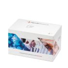

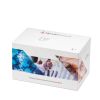
FH/FHR-1, Human, ELISA kit, 1 x 96 det. - HK342-01

Verwandte Produkte

The complement system is a key component of the immune response that defends against infections, initiates inflammation, manages immune complexes and regulation of immune responses. Complement factor H (CFH or FH) is the principal regulator of the alternative pathway, ensuring that complement activation is restricted to appropriate targets. Unlike the classical and lectin pathways, the alternative pathway is constitutively active at a low level and therefore requires tight control. CFH exerts this control by binding to C3b, accelerating decay of the C3 convertase (C3bBb), and acting as a cofactor for Complement factor I-mediated cleavage of C3b. In addition, CFH inhibits convertase formation by competing with factor B. Through these mechanisms, it regulates complement both in fluid phase and on host cell surfaces.
Structurally, CFH is a ~150 kDa plasma glycoprotein composed of linearly arranged 20 short consensus repeat (SCR) domains (also called complement control protein (CCP) modules). These domains confer multiple ligand-binding sites, including three regions for C3b and several for polyanions such as glycosaminoglycans. Functionally, CFH is organized into two main regions: the N-terminal SCR1–4 domains mediate complement regulatory activity, while the C-terminal domains, particularly SCR19–20 with contribution from SCR7, enable recognition of host surfaces via binding to C3 fragments and polyanions. This dual architecture allows CFH to selectively protect self-tissues from complement-mediated damage.
CFH is part of a protein family that includes the complement factor H-related (FHR) proteins (FHR1–FHR5), which share a similar SCR-based structure and high sequence homology, particularly in domains corresponding to CFH surface-recognition regions. Unlike FHL1, FHR1-5 proteins lack domains equivalent to CFH SCR1–4 and therefore generally lack intrinsic complement regulatory activity. Instead, they are thought to modulate complement by competing with CFH for ligand binding.
Complement factor H-related protein 1 shows the closest similarity to CFH. Its C-terminal domains are highly homologous to CFH SCR19–20, enabling binding to similar ligands such as C3b and polyanions. However, lacking regulatory domains, FHR1 can displace CFH from surfaces without inhibiting complement activation, potentially promoting local amplification. This competitive interaction is enhanced by FHR1 dimerization.
Dysregulation of the CFH–FHR balance is associated with diseases including Atypical hemolytic uremic syndrome, C3 glomerulopathy (aHUS), and age-related macular degeneration (AMD), highlighting the clinical importance of this regulatory network.
| Datasheet URL | https://www.hycultbiotech.com/wp-content/uploads/2022/06/HK342-0526.pdf |
|---|---|
| Quantity | 1x96det. |
| Quantity | 1 x 96 det. |
| Standard range | 3.9 - 250 ng/ml |
| Detection level | 3.9 ng/ml |
| Working volume | 100 μl/well |
| Species | Human |
| Application | Immuno assays |
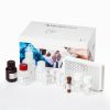
| Application | The human complement factor H ELISA kit is to be used for the in vitro quantitative determination of human complement factor H in serum, plasma and urine samples. |
| Principle | The human complement factor H ELISA is a ready-to-use solid-phase enzyme-linked immunosorbent assay based on the sandwich principle with a working time of 2½ hours. The efficient format of a plate with twelve disposable 8-well strips allows free choice of batch size for the assay. Samples and standards are incubated in microtiter wells coated with antibodies recognizing human complement factor H. Peroxidase conjugate antibody will bind to the captured human complement factor H. Peroxidase conjugate antibody will react with the substrate, tetramethylbenzidine (TMB). The enzyme reaction is stopped by the addition of oxalic acid. The absorbance at 450 nm is measured with a spectrophotometer. A standard curve is obtained by plotting the absorbance (linear) versus the corresponding concentrations of the human complement factor H standards (log). The human complement factor H concentration of samples, which are run concurrently with the standards, can be determined from the standard curve. |
| Storage and stability | Product should be stored at 4°C. Under recommended storage conditions, product is stable for at least six months. |
| Precautions | For research use only. Not for use in or on humans or animals or for diagnostics. It is the responsibility of the user to comply with all local/state and Federal rules in the use of this product. Hycult Biotech is not responsible for any patent infringements that might result with the use of or derivation of this product. |
| References | 1. Oppermann, M. et al; Quantitation of components of the alternative pathway of
complement (APC) by enzyme-linked immunosorbent assays. J Immunol Methods 1990,
133: 181 2. Oppermann M. et al; Elevated plasma levels of the immunosuppressive complement fragment Ba in renal failure. Kidney Int 1991, 40: 939 3. Pickering M. et al: Renal diseases associated with complement factor H: novel insights from humans and animals. Clin Exp Immunol 1993, 151: 210 4. Scambi, C et al; Comparative Proteomic Analysis of Serum from Patients with Systemic Sclerosis and Sclerodermatous GVHD. Evidence of Defective Function of Factor H. PlosOne 2010, 5:e12162 5. Brandstätter, H et al; Purification and biochemical characterization of functional complement factor H from human plasma fractions. Vox Sanguinis 2012 |
| Disease | Autoimmunity, Nephrology |
| Application: | Immuno assays |
|---|---|
| Application assays: | The human complement factor H ELISA kit is to be used for the in vitro quantitative determination of human complement factor H in serum, plasma and urine samples. |
| Principle: | The human complement factor H ELISA is a ready-to-use solid-phase enzyme-linked immunosorbent assay based on the sandwich principle with a working time of 2½ hours. The efficient format of a plate with twelve disposable 8-well strips allows free choice of batch size for the assay. Samples and standards are incubated in microtiter wells coated with antibodies recognizing human complement factor H. Peroxidase conjugate antibody will bind to the captured human complement factor H. Peroxidase conjugate antibody will react with the substrate, tetramethylbenzidine (TMB). The enzyme reaction is stopped by the addition of oxalic acid. The absorbance at 450 nm is measured with a spectrophotometer. A standard curve is obtained by plotting the absorbance (linear) versus the corresponding concentrations of the human complement factor H standards (log). The human complement factor H concentration of samples, which are run concurrently with the standards, can be determined from the standard curve. |
| Recovery: | Normal human blood samples (plasma), containing baseline levels of human complement factor H, were spiked with human complement factor H in concentrations of 62.5 and 15.6 ng/ml. Samples with and without human complement factor H were incubated for 1 hour at room temperature. Samples were measured using the ELISA. Values for human complement factor H ranged between 65% and 134% (mean 102%). |
Diese Artikel könnten Ihnen eventuell auch gefallen!