
Cyto3D Live-Dead Assay Kit (1ml)
Live dead cell viability analysis for 3D and 2D cell culture
Catalog #
BM01
313,00 €

The Cyto3D™ Live-Dead Assay Kit is used to determine the live/dead nucleated cells by using a fast one-step staining procedure for analysis on a dual-fluorescence system. This kit is recommended for viability analysis of cells cultured in 3D, 2D coating and on monolayer.
- Ready-to-use
- Fast
- Sensitive
- Excellent for 3D cell cultures
- Cost-effective
Acridine orange (AO) and propidium iodide (PI), both nuclear staining (nucleic acid binding) dyes, are used in this kit. AO is permeable to both live and dead cells and stains all nucleated cells to generate green fluorescence. PI only penetrates the membranes of nucleated cells with compromised membranes and stains the dead cells to generate red fluorescence. Due to the quenching, when cells are stained with both AO and PI, all live nucleated cells fluoresce green and all dead nucleated cells fluoresce red (the PI reduces the fluorescence intensity of the AO by fluorescence resonance energy transfer (FRET)). Non-nucleated materials such as red blood cells, platelets and debris do not fluorescence and are ignored by fluorescence microscopes.
Dual-Fluorescence Viability, using AO and PI, is the recommended viability analysis method for cell lines, primary cells, and stem cells.
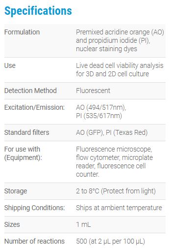
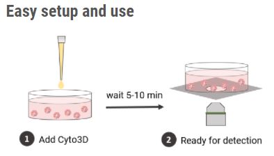
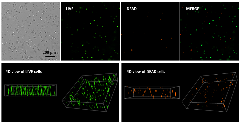
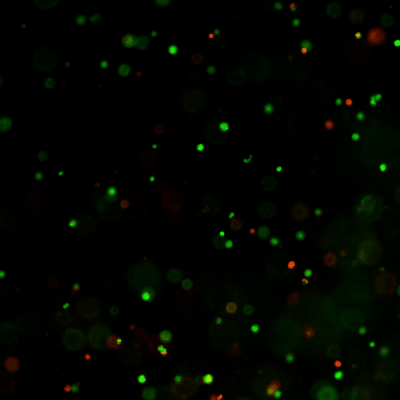

Figure 1. Live-dead cell viability analysis by using Cyto3D Live-Dead Assay Kit.
Glioblastoma cells (SF 298, about 60% cell viability) were 3D cultured in VitroGel system for 2 days. 2 µL of Cyto3D reagent was added to each well containing 50 µL hydrogel and 50 µL cover medium. The mixture was incubated at 37 °C for 5-10 min. The cells were then observed under a fluorescent microscope. The images show the Live (green) and Dead (orange) cells within the 3D hydrogel matrix. The z-stack images of cells within hydrogel were then 3D reconstructed and showed in the 4D view images. The live and dead cells at higher levels of the hydrogel clearly show in the images by using Cyto3D Live-Dead Assay Kit.
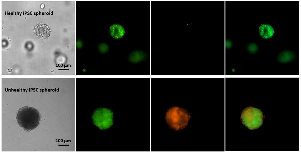
Figure 2. Live-dead cell viability images of stem cell spheroids.
Stem cells were static suspension-cultured in VitroGel STEM (CAT# VHM02) for 5 days. 2 µL of Cyto3D reagent was added to each well containing 100 µL cell suspension. The mixture was incubated at 37 °C for 5-10 min. The cells were then observed under a fluorescent microscope. The images show the Live (green) and Dead (orange) stem cell spheroids cultured in a 3D hydrogel matrix. The live-dead dyes of Cyto3D Live-Dead Assay Kit can successfully penetrate into large cell spheroids for cell viability analysis.
References/Publications
- Powell K. Adding depth to cell culture. Science, 356(6333), 96–98. https://doi.org/10.1126/science.356.6333.96
- Ready-to-use
- Fast
- Sensitive
- Excellent for 3D cell cultures
- Cost-effective
Acridine orange (AO) and propidium iodide (PI), both nuclear staining (nucleic acid binding) dyes, are used in this kit. AO is permeable to both live and dead cells and stains all nucleated cells to generate green fluorescence. PI only penetrates the membranes of nucleated cells with compromised membranes and stains the dead cells to generate red fluorescence. Due to the quenching, when cells are stained with both AO and PI, all live nucleated cells fluoresce green and all dead nucleated cells fluoresce red (the PI reduces the fluorescence intensity of the AO by fluorescence resonance energy transfer (FRET)). Non-nucleated materials such as red blood cells, platelets and debris do not fluorescence and are ignored by fluorescence microscopes.
Dual-Fluorescence Viability, using AO and PI, is the recommended viability analysis method for cell lines, primary cells, and stem cells.
Figure 1. Live-dead cell viability analysis by using Cyto3D Live-Dead Assay Kit.
Glioblastoma cells (SF 298, about 60% cell viability) were 3D cultured in VitroGel system for 2 days. 2 µL of Cyto3D reagent was added to each well containing 50 µL hydrogel and 50 µL cover medium. The mixture was incubated at 37 °C for 5-10 min. The cells were then observed under a fluorescent microscope. The images show the Live (green) and Dead (orange) cells within the 3D hydrogel matrix. The z-stack images of cells within hydrogel were then 3D reconstructed and showed in the 4D view images. The live and dead cells at higher levels of the hydrogel clearly show in the images by using Cyto3D Live-Dead Assay Kit.
Figure 2. Live-dead cell viability images of stem cell spheroids.
Stem cells were static suspension-cultured in VitroGel STEM (CAT# VHM02) for 5 days. 2 µL of Cyto3D reagent was added to each well containing 100 µL cell suspension. The mixture was incubated at 37 °C for 5-10 min. The cells were then observed under a fluorescent microscope. The images show the Live (green) and Dead (orange) stem cell spheroids cultured in a 3D hydrogel matrix. The live-dead dyes of Cyto3D Live-Dead Assay Kit can successfully penetrate into large cell spheroids for cell viability analysis.
References/Publications
- Powell K. Adding depth to cell culture. Science, 356(6333), 96–98. https://doi.org/10.1126/science.356.6333.96
| MCE Cat. No. | BM01 |
|---|---|
| Datasheet URL | https://cdn.thewellbio.com/wp-content/uploads/2020/11/Cyto3D-Live-Dead-Assay-Kit-TDS.pdf |
| Quantity | 1 ml |